PNH Emergency Triage: Visceral Thrombosis (Budd-Chiari) & Breakthrough Hemolysis (BTH)
Most emergency room clinicians see fewer than one case of Paroxysmal Nocturnal Hemoglobinuria in a decade. When a PNH patient arrives at an emergency department at 2 AM with acute upper abdominal pain, severe headache, or dark cola-colored urine during a viral infection, standard ER triage protocols can miss the two life-threatening complications of PNH: visceral/cerebral venous thrombosis and complement-amplified breakthrough hemolysis (BTH).
Bookmark or print this page to hand directly to the triage nurse and attending emergency physician.
Venous thromboembolism is the leading cause of mortality in untreated PNH, striking unusual anatomical sites including the hepatic veins (Budd-Chiari syndrome), portal and mesenteric veins, and cerebral dural venous sinuses. Any PNH patient presenting with acute abdominal pain, severe headache, or infection-triggered dark urine requires urgent imaging, therapeutic anticoagulation, and immediate complement blockade.
1. Disease Mechanism: This patient has Paroxysmal Nocturnal Hemoglobinuria (PNH), a complement-mediated disorder with extreme hypercoagulability and intravascular hemolysis.
2. If Presenting With Fever (>=38.0°C / 100.4°F): Complement inhibitors (ravulizumab, eculizumab, pegcetacoplan, iptacopan, danicopan, crovalimab) block bactericidal immunity against Neisseria meningitidis and encapsulated bacteria. Draw blood cultures and administer IV ceftriaxone (2 g IV) immediately before waiting for lumbar puncture or imaging.
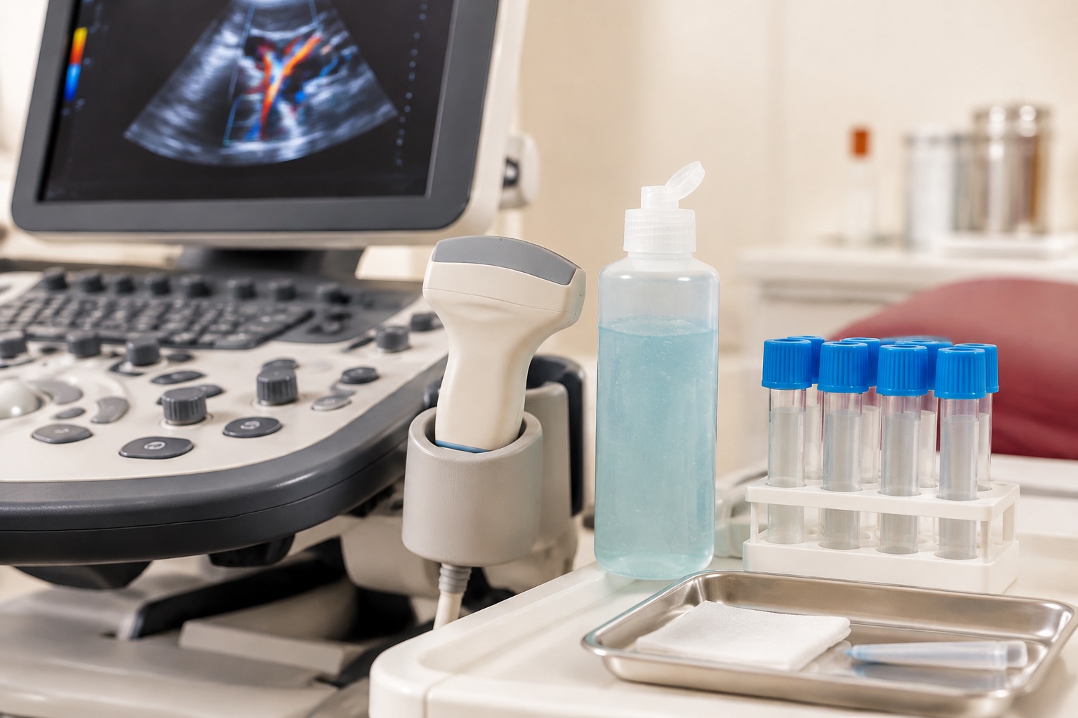
3. If Presenting With Abdominal Pain or Severe Headache: Rule out Budd-Chiari syndrome (hepatic vein thrombosis), portal/mesenteric vein thrombosis, and cerebral venous sinus thrombosis (CVST) via Doppler ultrasound, contrast CT venography, or MR venography. Note: Platelets may be low-normal due to concurrent marrow failure; do not rely on D-dimer alone.
4. Never Hold Complement Inhibitors: Do NOT discontinue oral iptacopan, danicopan, subcutaneous pegcetacoplan, or scheduled IV C5 infusions during hospitalization. Stopping therapy triggers catastrophic rebound hemolysis and thrombosis. Contact the patient's on-call hematologist immediately.
Why Does PNH Cause Blood Clots in the Liver and Brain Veins Instead of Just the Legs?
In untreated PNH, C5a anaphylatoxin release, nitric oxide depletion, platelet microparticle shedding (from CD59-deficient platelets), and impaired fibrinolysis (loss of GPI-anchored urokinase plasminogen activator receptor, uPAR) create a prothrombotic storm that preferentially targets low-flow splanchnic and cerebral venous beds:
| Thrombotic Site & Share of PNH Clots | Warning Symptoms | First-Line Diagnostic Imaging | Acute Hematology Management |
|---|---|---|---|
| Hepatic Veins (Budd-Chiari Syndrome) ~25% to 40% of all PNH thromboses |
Acute or subacute right upper quadrant abdominal pain, rapid abdominal distension (ascites), tender hepatomegaly, jaundice, elevated AST/ALT. | Color Doppler Ultrasound of hepatic veins & IVC, confirmed by Triple-Phase Contrast CT or MRI. | Immediate therapeutic anticoagulation (UFH or LMWH) plus urgent initiation of terminal C5 inhibition (eculizumab/ravulizumab) to halt ongoing clot propagation; avoid unnecessary liver biopsy. |
| Portal, Mesenteric & Splenic Veins ~15% to 20% of PNH thromboses |
Severe colicky mid-abdominal pain out of proportion to physical exam, nausea, vomiting, fever, or occult gastrointestinal bleeding. | Contrast-Enhanced Abdominal CT (Portal Venous Phase); see also CT Renal Hemosiderosis. | Immediate IV anticoagulation + complement blockade. Note: Smooth muscle dystonia from free Hb also causes abdominal pain, so imaging is mandatory to distinguish spasm from ischemia. |
| Cerebral Dural Venous Sinuses (CVST) ~10% to 15% of PNH thromboses |
New, severe, unrelenting headache (often worse when lying flat or coughing), blurred vision, papilledema, focal seizures, or focal neurological deficits. | MR Venography (MRV) or CT Venography (CTV) (plain non-contrast head CT misses up to 30% of CVST cases!). | Urgent therapeutic LMWH/UFH (even in the presence of small venous hemorrhagic infarction, per AHA/ESO CVST guidelines) + immediate C5 inhibitor loading. |
| Deep Vein Thrombosis (DVT), PE & Dermal Veins ~25% to 35% of PNH thromboses |
Unilateral leg swelling/pain, acute dyspnea/pleuritic chest pain, or painful violaceous skin plaques (dermal vein thrombosis). | Compression venous duplex ultrasound; CT pulmonary angiography (CTPA) for PE (with adequate IV hydration to protect kidneys). | Anticoagulation alone has a high recurrence rate in untreated PNH; complement inhibition is the primary anti-thrombotic therapy. |
Pharmacokinetic (PK) vs. Pharmacodynamic (PD) Breakthrough Hemolysis: How to Tell Them Apart
Breakthrough hemolysis (BTH) is defined as the sudden return of intravascular hemolysis (dark urine, dysphagia, severe fatigue, hemoglobin drop >=2 g/dL, and LDH spiking >2x ULN) in a patient previously controlled on a complement inhibitor. Hematologists classify BTH into two distinct categories:
1. Pharmacokinetic (PK) Breakthrough Hemolysis
LOW DRUG TROUGH LEVELTiming: Occurs predictably in the final 24 to 72 hours before the next scheduled infusion (e.g., Day 12-14 on eculizumab) or after a missed oral/subcutaneous dose.
Mechanism: Free drug concentration falls below the threshold needed to neutralize baseline complement turnover (50 ug/mL for eculizumab; 100 ug/mL for ravulizumab).
Solution: If on eculizumab 900 mg q2w, either switch to ravulizumab (Ultomiris) q8w (which eliminates PK troughs) or increase eculizumab to 1,200 mg q2w. If on oral iptacopan or SC pegcetacoplan, take the missed dose immediately and reinforce dosing alarms.
2. Pharmacodynamic (PD) Breakthrough Hemolysis
COMPLEMENT AMPLIFYING CONDITION (CAC)Timing: Occurs at any point in the dosing cycle, triggered by an acute Complement-Amplifying Condition (CAC): bacterial/viral infection (COVID-19, influenza, gastroenteritis, UTI), major surgery, trauma, or third-trimester pregnancy.
Mechanism: Massive classical and lectin pathway activation generates high-density C3b surface clusters that overwhelm standard complement inhibitor blockade.
Solution: Treat the underlying infection aggressively, maintain hydration, and on proximal inhibitors (pegcetacoplan/iptacopan) with severe BTH (LDH >3x-5x ULN), administer short-term intensive dosing (e.g., daily SC pegcetacoplan x3 days or a single rescue dose of IV eculizumab/ravulizumab) per specialist protocol.
Frequently Asked Hematology & Patient Questions
Direct, link-free clinical answers grounded in ASH, IPIG, and Phase 3 complement inhibitor trial evidence.
Do PNH patients need lifelong blood thinners (anticoagulation) if they are on Ultomiris or Soliris?
In PNH patients who have never had a blood clot (primary prophylaxis), starting effective complement inhibition (ravulizumab, eculizumab, pegcetacoplan, or iptacopan) reduces thrombotic risk so effectively that routine lifelong anticoagulation is generally not required unless other risk factors exist. In patients who have already suffered a prior PNH-related thrombosis (secondary prophylaxis), anticoagulation is continued alongside complement blockade, though selected patients achieving durable complete hemolytic control without anatomical vein damage may eventually taper anticoagulation under specialist supervision.
Clinical source: Hillmen et al., Blood 2007; Griffin et al., Br J Haematol 2021.
Why must red blood cell transfusions in PNH use leukoreduced packed red cells?
Historically, old medical textbooks recommended saline-washed red blood cells for PNH to avoid transfusing donor plasma complement. Modern third-generation leukoreduced packed red blood cells contain negligible donor plasma and can be safely transfused without saline washing, while preventing febrile non-hemolytic transfusion reactions (which release inflammatory cytokines that can amplify complement activation).
Clinical source: Parker et al., Blood 2005 · See Perioperative Guide.
Can dehydration or strenuous marathon exercise trigger a PNH hemolytic flare?
Yes. Severe dehydration, prolonged endurance exercise (which causes transient lactic acidosis and mechanical foot-strike hemolysis), excessive alcohol binges, and viral gastroenteritis lower local microcirculatory pH and activate the alternative complement pathway. Maintaining steady hydration and electrolytes helps buffer against mild non-infectious flares.
Clinical source: AAMDSIF Clinical Guide · See Living With PNH.