PNH Flow Cytometry Decoder (CD55, CD59, FLAER) & CT Imaging Signs
When patients open their laboratory portal after a bone marrow failure or hemolytic anemia workup, they are confronted with a table of unfamiliar percentages: Granulocyte FLAER/CD24 deficiency, Monocyte FLAER/CD14 deficiency, and RBC CD59 Type I, Type II, and Type III populations.
At the same time, a substantial number of PNH patients are first suspected not in a hematology clinic, but in a radiology suite after an abdominal CT or MRI ordered for abdominal pain or dark urine reveals either visceral venous thrombosis or renal cortical hemosiderosis.
High-sensitivity peripheral blood flow cytometry using FLAER and monoclonal antibodies against CD55, CD59, CD24, and CD14 is the gold standard for diagnosing PNH. Granulocyte and monocyte clone percentages reflect true bone marrow PNH stem cell burden, whereas red blood cell clone percentages are frequently underestimated due to hemolysis and blood transfusions.
Why Is My Granulocyte PNH Clone 72% While My Red Blood Cell Clone Is Only 21%?
This is the single most common question on PNH patient forums. An acquired PIGA mutation occurs in a multipotent hematopoietic stem cell in the bone marrow, meaning the bone marrow produces the exact same proportion of GPI-deficient neutrophils, monocytes, and red blood cells.
However, once those cells enter peripheral blood, their lifespans diverge dramatically:
- Neutrophils (Granulocytes) & Monocytes Survive Normally: Even without CD55 and CD59, white blood cells have nucleated membrane repair mechanisms and a short 6-to-24-hour circulation half-life. They are not lysed by complement and are never diluted by red cell transfusions. Therefore, the granulocyte FLAER/CD24-deficient percentage is the true benchmark of your PNH clone size.
- Type III Red Blood Cells Are Destroyed in Circulation: Normal red blood cells live 120 days, whereas untreated Type III PNH red blood cells (completely lacking CD59) survive only 3.5 to 10 days before being lysed by the C5b-9 Membrane Attack Complex. Furthermore, packed red blood cell transfusions add normal (Type I) donor cells, artificially depressing the measured RBC PNH clone percentage.
- What Happens After Starting a Complement Inhibitor: Once you start ravulizumab, eculizumab, pegcetacoplan, or iptacopan, your Type III red blood cells are protected from destruction. Over 8 to 16 weeks, you will see your RBC PNH clone percentage climb to match your granulocyte clone percentage. A rising RBC clone percentage after starting therapy means the drug is working to keep your red cells alive, not that the disease is worsening!
| Laboratory Assay | Target / Reagent | Diagnostic Threshold | Clinical Pitfalls & Confounders |
|---|---|---|---|
| Granulocyte Flow Cytometry CD15+ gated neutrophils |
FLAER + CD24 (or CD157) | >=0.01% (High-sensitivity); >=10% typical in hemolytic PNH | Never affected by RBC transfusions. Gold standard for tracking clone expansion or remission over time. |
| Monocyte Flow Cytometry CD64+ / CD33+ gated monocytes |
FLAER + CD14 (or CD157) | Corroborates granulocyte clone (>=0.01%) | Diagnosing PNH requires demonstrating GPI deficiency in at least two distinct blood lineages (e.g., granulocytes + monocytes or RBCs) to rule out rare congenital single-protein deficiencies. |
| Erythrocyte (RBC) Flow Cytometry CD235a (Glycophorin A) gated |
Anti-CD59 + Anti-CD55 | Separates Type I, Type II, and Type III populations | FLAER cannot be used on red blood cells because human erythrocytes lack the specific surface receptors for aerolysin binding; anti-CD59 monoclonal antibody is mandatory. |
| Lactate Dehydrogenase (LDH) Serum enzyme |
Ratio vs. Lab Upper Limit of Normal (ULN) | >=1.5x ULN defines clinically significant intravascular hemolysis | In untreated classic PNH, LDH often reaches 4x to 10x ULN. An LDH >=1.5x ULN combined with a large clone increases thromboembolic risk more than 7-fold. |
| Haptoglobin & Reticulocytes | Free Hb scavenger & marrow output | Haptoglobin <10 mg/dL (undetectable); Retics >100 x 10^9/L | If reticulocytes are low (<60 x 10^9/L) despite severe anemia and high LDH, investigate concurrent aplastic anemia marrow failure or folate/iron depletion. |
Can Paroxysmal Nocturnal Hemoglobinuria Be Suspected or Diagnosed on CT and MRI Scans?
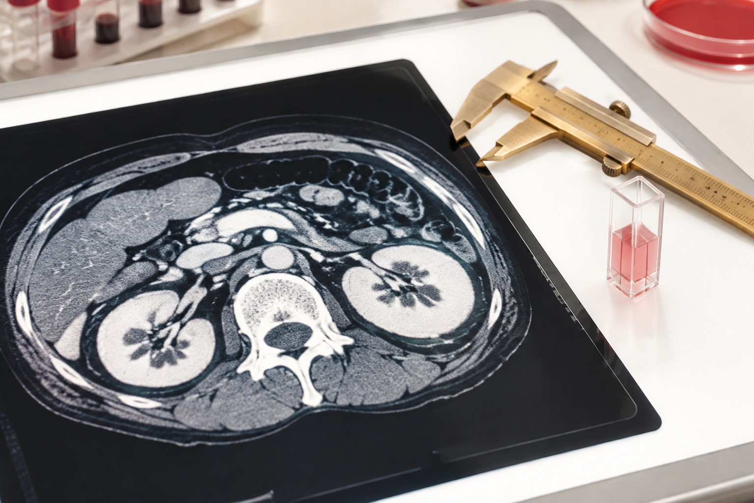
In historical literature and on the original PNHdisease.org clinical library (including H. Rochant's MRI monograph impact_of_mri.pdf and abdominal imaging dossier widespread_abdominal_venous.pdf), many PNH patients with unrecognized chronic hemolysis were first identified after undergoing abdominal CT or MRI for unexplained abdominal pain or hematuria/hemoglobinuria workups.
1. Unenhanced Abdominal CT: Hyperdense Renal Cortex
CT ATTENUATION >50-65 HUDuring chronic intravascular hemolysis, cell-free hemoglobin dimers pass through the renal glomeruli and are reabsorbed by the proximal convoluted tubules in the outer renal cortex. Once ferritin capacity is saturated, iron is stored as insoluble hemosiderin granules selectively in the renal cortex, sparing the renal medulla.
On a non-contrast (unenhanced) abdominal CT scan, this iron overload produces symmetrical, diffusely increased attenuation (bright density of 50 to 70 Hounsfield Units) of the bilateral renal cortices relative to the darker renal pyramids, a striking finding that radiologists recognize as renal cortical hemosiderosis.
2. Abdominal MRI (T1 / T2*): The Reversed Cortical Rim Sign
MRI T2* SUSCEPTIBILITYMagnetic resonance imaging is even more sensitive to paramagnetic hemosiderin iron in the renal cortex. On normal kidneys, T1-weighted MRI shows the renal cortex as brighter (higher signal) than the medulla. In PNH, paramagnetic hemosiderin causes profound T2* and T1 relaxation shortening, turning the outer renal cortex jet-black (profoundly hypointense) on both T1-weighted and T2-weighted images.
Crucially, unlike systemic hemochromatosis or transfusional iron overload (which deposits iron heavily in the liver and spleen parenchyma), untreated PNH shows selective renal cortical iron overload with relative sparing of the spleen until repeated transfusions or C5-inhibitor extravascular hemolysis occur.
When a patient presents to the emergency room with acute left or right upper quadrant abdominal pain, contrast-enhanced CT frequently reveals hepatic vein thrombosis (Budd-Chiari syndrome), portal vein thrombosis, or mesenteric/splenic vein thrombosis alongside high-attenuation renal cortices. International hematology guidelines mandate high-sensitivity PNH flow cytometry in every patient with unexplained splanchnic (intra-abdominal) or cerebral venous thrombosis. Read the Thrombosis & BTH Emergency Guide.
Frequently Asked Hematology & Patient Questions
Direct, link-free clinical answers grounded in ASH, IPIG, and Phase 3 complement inhibitor trial evidence.
Why is the Coombs test (Direct Antiglobulin Test) negative in newly diagnosed PNH?
The direct Coombs test (DAT) detects antibodies (IgG) or complement C3d bound to the surface of red blood cells. In untreated PNH, hemolysis is driven by an intrinsic deficiency of GPI-anchored CD55 and CD59 rather than autoantibodies, and C5b-9 lyses the cells so rapidly that C3d-coated cells do not accumulate. Thus, Coombs is negative at diagnosis. However, once a patient starts a C5 inhibitor (eculizumab or ravulizumab), PNH red cells survive longer and become coated with C3d, often turning the C3d Coombs test positive.
Clinical source: Risitano et al., Blood 2009 / 2010 · See C5 vs. Proximal Inhibitors.
Can a blood transfusion hide a PNH diagnosis on flow cytometry?
A recent packed red blood cell transfusion can significantly dilute the measured PNH clone percentage in erythrocytes (CD59 analysis) because transfused donor red cells are normal Type I cells. However, transfusions never affect neutrophil (granulocyte) or monocyte FLAER flow cytometry. Therefore, high-sensitivity flow cytometry on white blood cells accurately diagnoses PNH even immediately after multiple blood transfusions.
Clinical source: Borowitz et al., Cytometry Part B 2010.
What does renal cortical hemosiderosis on CT or MRI mean for my kidney function?
Renal cortical hemosiderosis on CT or MRI proves that your kidneys have been filtering cell-free hemoglobin from chronic intravascular hemolysis for months or years. Over time, tubular iron toxicity and nitric oxide scavenging can cause chronic kidney disease (CKD stages 1 to 3) and Fanconi-like tubular dysfunction. Starting effective complement inhibition halts further hemoglobinuria and allows renal cortical iron deposits to clear gradually over 12 to 24 months.
Clinical source: Hillmen et al., Am J Hematol 2010.